
Fig. 7.
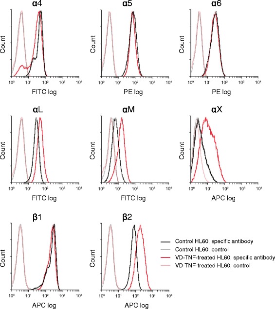
Cell surface expression of integrin subunit proteins in HL60 cells. Representative histograms from flow cytometric analyses, showing the cell surface expression of α4, α5, α6, αL, αM, αX, β1, and β2 integrin subunits in HL60 cells of the control group (black line, specific antibody; gray line, control) and in the VD-TNF group (red line, specific antibody; pink line, control). For the control in each group, cell suspensions were pretreated with the human Fc receptor-binding inhibitor and with an APC-conjugated isotype control antibody in case of the APC-conjugated anti-αX, anti-β1, and anti-β2 antibody. All integrin subunits are clearly expressed except for the αX integrin subunit in HL60 cells of the control group