Fig. 2.

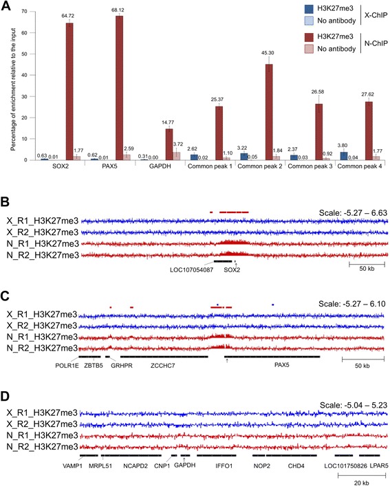
N-ChIP and X-ChIP results at control loci. a Enrichment of H3K27me3 and no antibody relative to the input measured by qPCR at three control gene loci: SOX2 (chr9: 16,918,111–16,919,468), PAX5 (chrZ: 81,789,479–81,896,738) and GAPDH (chr1: 76,950,864–76,956,805), and at the localization of common enrichment detected with epic (see Fig. 1 legend for peak description). H3K27me3 enrichments are represented in blue for X-ChIP and in red for N-ChIP, no-antibody enrichments are represented in light blue for X-ChIP and in light red for N-ChIP. Error bars: SEM. b-d Visualization with IGV of H3K27me3 enrichment normalized to input [log2(IP/input)]. X-ChIP-seq muscle tracks are shown in blue, N-ChIP muscle tracks are shown in red. The colored boxes above the tracks represent broad peaks detected by epic in either X-ChIP-seq (blue) or N-ChIP muscle (red) replicates. The black arrows under the tracks represent the position of qPCR primers at (b) SOX2, (c) PAX5 and (d) GAPDH
