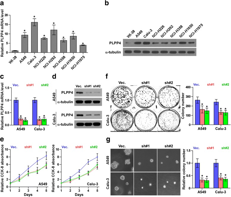
Fig. 4.

Silencing PLPP4 inhibits proliferation in lung carcinoma cells. a and b Real-time PCR and Western blotting analysis of PLPP4 expression in WI-38 and lung carcinoma cell lines. GAPDH was used as the endogenous control for RT-PCR and α-Tubulin was used as the loading control in the Western blot. Each bar represents the mean values ± SD of three independent experiments. *P < 0.05. c and d Real-time PCR and Western blot of the indicated lung carcinoma cells transfected with PLPP4-RNAi-vector, PLPP4-RNAi#1 and PLPP4-RNAi#2. GAPDH was used as the endogenous control for RT-PCR and α-Tubulin was used as the loading control in the Western blot. Each bar represents the mean values ± SD of three independent experiments. *P < 0.05. e CCK8 assays revealed that silencing PLPP4 reduced cell viability in lung carcinoma cells. Each bar represents the mean values ± SD of three independent experiments. *P < 0.05. f Silencing PLPP4 reduced the mean colony number according to the colony formation assay. Each bar represents the mean values ± SD of three independent experiments. *P < 0.05. g Representative micrographs and colony numbers from the anchorage-independent growth assay. Each bar represents the mean values ± SD of three independent experiments. *P < 0.05
