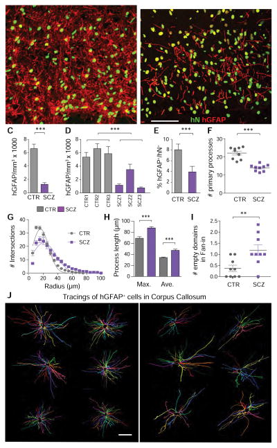
Figure 3. Astrocytic differentiation is impaired in schizophrenia hGPC chimeric brain.
Human iPSC GPC chimeras were established in immunodeficient shiverer hosts and sacrificed at 19 weeks, and astrocytic diffeerntiation assessed. A–B, representative images of the corpus callosum of mice neonatally injected with iPSC GPCs derived from either control (A, line 22) or schizophrenic (B, line 164) subjects (human nuclear antigen, green; glial fibrillary acidic protein, red). A, Control hiPSC GPCs from all tested patients rapidly differentiated as GFAP+ astrocytes with dense fiber arrays in both callosal white and cortical gray matter. B, In contrast, SCZ GPCs were slow to develop mature GFAP expression. At 19 weeks, GFAP+ astrocyte densities were significantly greater in mice chimerized with control than SCZ-derived GPCs, both as a group (C), and when analyzed line-by-line (D). This was not just a function of less callosal engraftment, as the proportion of human donor cells that developed GFAP and astrocytic phenotype was significantly lower in SCZ- than control GPC-engrafted mice (E). Sholl analysis of individual astroglial morphologies(Sholl, 1953), as imaged in 150 μm sections and reconstructed in 3D by Neurolucida (J), revealed that astrocytes in SCZ hGPC chimeras differed significantly from their control hGPC-derived counterparts, with fewer primary processes (F), less proximal branching (G), and longer distal fibers (H). When the 3-D tracings (J) were assessed by Fan-in radial analysis (MBF Biosciences)(Dang et al., 2014), control astrocytic processes were noted to extended uniformly in all directions, but SCZ astrocyte processes left empty spaces, indicative of a discontiguous domain structure (I). ***p<0.0001, by t-test (C, E, F, H; by 2-way ANOVA in D; **p<0.002 in I; p<0.0001 by non-linear comparison in G.
Scale, A–B = 50 μm, J = 25 μm.

