
Figure 3. Microscopic visualization of the gut microbiota.
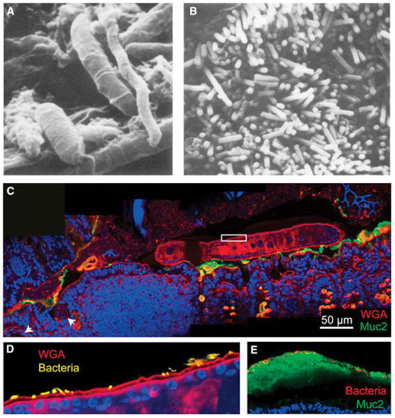
A) Scanning electron micrograph (Savage and Blumershine, 1974) from the mouse distal gut. Rod-, fusiform- and spiral-shaped bacteria are present, illustrating the morphological diversity of the gut microbiota.
B) Scanning electron micrograph from the mouse colon (Savage and Blumershine, 1974), highlighting the high density of bacteria in the gut.
C) Inter-kingdom spatial interactions between helminths and bacteria have been poorly studied to date. Trichuris muris, a mouse model of whipworm, was visualized by labeling with WGA (red) in the proximal colon of a conventional Swiss-Webster mouse 14 days after inoculation with ~200 T. muris ova. The anterior end is embedded in the epithelium near gastrointestinal lymphoid tissue (white arrowheads), while the posterior end is free in the lumen. MUC2 is labeled in green, and the mouse and worm nuclei are labeled with DAPI in blue.
D) Magnification of white box in (C) with bacterial DAPI signal segmented from the worm DAPI signal and false-colored yellow. Bacteria can be seen embedded in the cuticle of the worm.
E) Much of what we know about localization in the gut is derived from mouse studies. Further studies of human biopsies are needed, and in particular, on samples with preserved mucus. Here, a biopsy from a healthy patient has been fixed in methacarn, processed, sectioned and stained as described in Box 1. Bacteria, which are labeled with DAPI and false-colored red, are visible on the luminal side of the inner mucus layer.