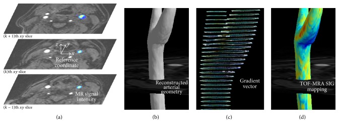
Figure 2.
Visualization and TOF-MRA signal intensity gradient (SIG). (a) The reference coordinate settings on TOF-MRA axial source images and the selected (left carotid) arterial color display. (b) 3D reconstruction of arterial geometry using the arterial threshold value. (c) A gradient vector setting: from the reference point on the artery wall (contour line), the position of inner point is identified with both a specific distance (0.03 mm in the present study) and the direction having the maximum gradient of the signal intensity. For the drawing, the gradient vector is lengthened to 0.3 mm. (d) 3D mapping of the carotid arterial TOF-MRA SIG.

