Figure 1.
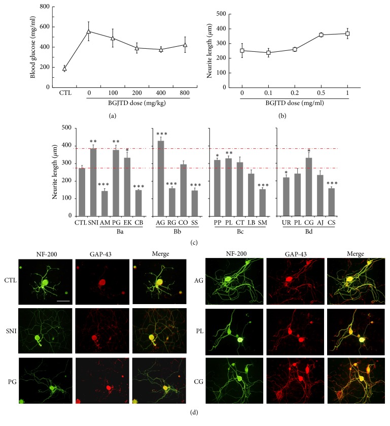
Dose responses to BGJTD and comparison of neurite outgrowth in DRG neurons after the treatment of individual constituents of BGJTD. (a) Changes of blood glucose levels with increasing doses of BGJTD. DGJTD up to 800 mg/kg was administered for 7 days to STZ-diabetic mice. Number of animals per group = 4. (b) Measurement of neurite length of DRG neurons in culture after the treatment of BGJTD. DRG neurons prepared from rats were cultured for 48 h in the presence of BGJTD at 0–1.0 mg/ml, and neurite length was measured. Number of independent experiments = 4. (c) Comparison of neurite outgrowth in individual subgroups Ba–Bd. Upper and lower red dotted lines denote the values of neurite length from animals given preconditioning nerve injury (SNI) and untreated control (CTL), respectively. Mean values of neurite length were compared among experimental groups (number of independent experiments = 4). ∗p < 0.05, ∗∗p < 0.01, ∗∗∗p < 0.001 versus untreated control (CTL). Unabbreviated names of all herbal drugs are described in Materials and Methods. (d) Representative fluorescence images of DRG neurons treated with herbal drugs scoring the highest neurite extension from each subgroup, along with CTL and SNI groups. In (c) and (d), herbal drugs (0.5 mg/ml) were treated to DRG neurons for 48 h and harvested for immunofluorescence staining for NF-200 (green) and GAP-43 (red) signals. Scale bar in (d) = 100 μm.

