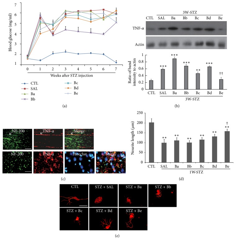
Figure 2.
Effects of treatment of subgroup decoctions on blood glucose levels, TNF-α production, and neurite outgrowth. (a) A time-dependent profile of blood glucose levels in different subgroups for 7 weeks after STZ injection. Number of animals in each group = 5. (b) Western blot analysis of TNF-α. Images in the upper panel show the representatives from 3 independent experiments, and quantitation of protein band intensity relative to actin control is shown in the lower panel. ∗∗p < 0.01, ∗∗∗p < 0.001 versus untreated control (CTL); ††p < 0.01 versus saline control. (c) Representative immunofluorescence images showing TNF-α and NF-200 signals in the sciatic nerve sections prepared from STZ-diabetic animals. Arrows in the upper panel indicate TNF-α signals merged with NF-200-stained axons in longitudinal sections. TNF-α signals were also found around the Hoechst-stained nuclei (arrows in lower panel, transverse section). The sciatic nerves for western analysis in (b) were prepared from 3W-STZ animals treated with subgroup drugs as indicated in the figure, and the nerves for immunostaining in (c) were from 3W-STZ animals with saline injection. (d) Comparison of neurite outgrowth in DRG neurons prepared from STZ-diabetic animals. ∗∗p < 0.01 versus untreated control; †P < 0.05 versus saline control. Number of independent experiments = 4. (e) Representative images of immunofluorescence staining of DRG neurons with NF-200. In (d) and (e), 7 days after STZ injection, DRGs at lumbar levels 4 and 5 were collected from animals showing blood glucose levels higher than 3 mg/ml and used for DRG neuron culture. Cells were treated with herbal drugs (0.5 mg/ml) for 48 h before the harvest of cells for immunofluorescence staining with NF-200 (red in (e)). Scale bars in (c) and (e) = 100 μm.

