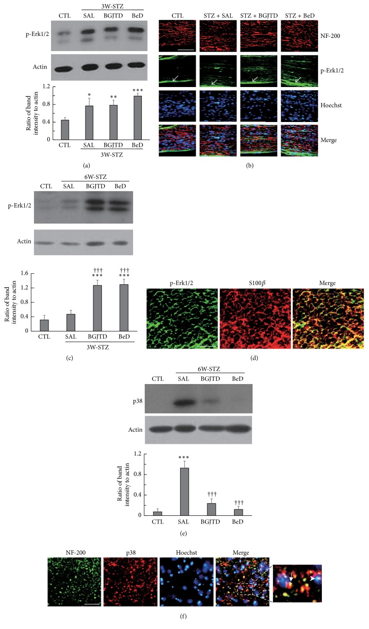
Figure 4.
Induction pattern of phospho-Erk1/2 and p38 proteins in the sciatic nerve of STZ-diabetic animals after herbal drug treatments. Animals were injected with STZ and, 1 week and 4 weeks later, subjected to daily administration of herbal drugs for 2 weeks (labeled 3W-STZ and 6W-STZ respectively). (a) Western blot analysis of phospho-Erk1/2 in the sciatic nerve from 3W-STZ animal group. (b) Immunofluorescence images showing phospho-Erk1/2 signals in NF-200-stained sciatic nerve sections, which were prepared from BeD-treated 3W-STZ animals. Phospho-ERK1/2 signals in the epineurial sheath are indicated by arrows. (c) Western blot analysis of phospho-Erk1/2 in the sciatic nerve from 6W-STZ animal group. (d) Immunofluorescence images showing phospho-Erk1/2 and S100β signals in the transverse nerve section, which were prepared from BeD-treated 6W-STZ animals. (e) Western blot analysis of p38 in the sciatic nerve from 6W-STZ animal group. (f) Immunofluorescence images showing p38 and NF-200 signals in the transverse nerve section, which were prepared from saline-treated 6W-STZ animals. Western blotting images in (a), (c), and (e) are the representatives from 3 independent experiments (upper panels), and quantitation of protein band intensity relative to actin control is shown in the lower panels. ∗p < 0.01, ∗∗p < 0.01, ∗∗∗p < 0.001 versus untreated control (CTL); †††p < 0.001 versus saline control. Scale bars in (b), (d), and (f) = 100 μm.

