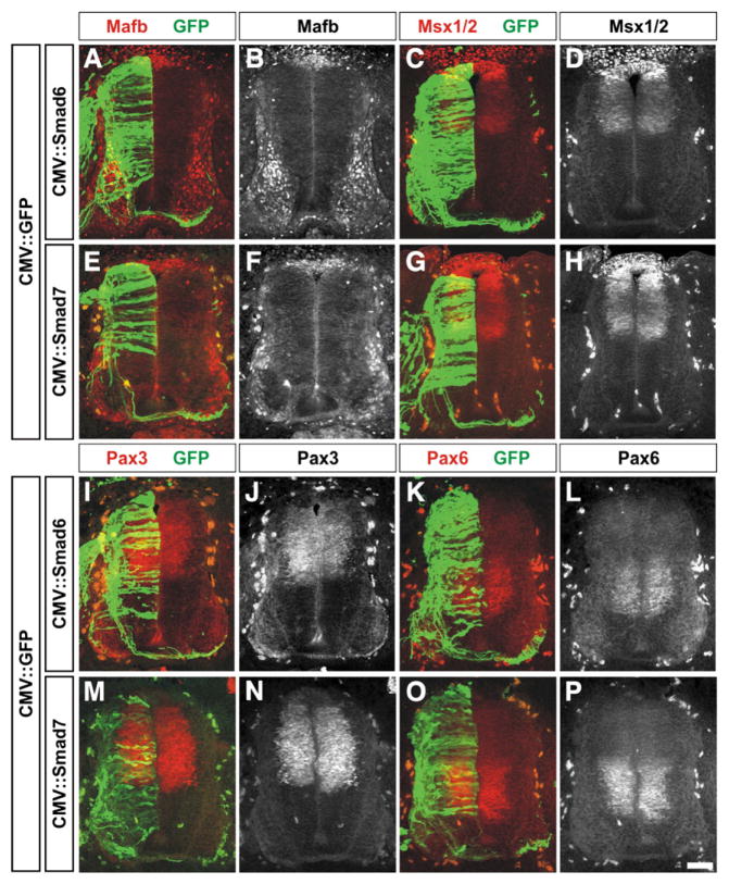
Fig. 3.
Mis-expression of Smad6 and Smad7 does not affect the distribution of many markers of the roof plate and dorsal and intermediate spinal cord. (A–P) GFP (green) in combination with either Smad6 (A–D and I–L) or Smad7 (E–H and M–P) were ectopically expressed throughout the spinal cord under the control of the CMV enhancer at HH stages 14/15. Embryos were harvested at HH stages 24/25 and examined for the distribution of markers that broadly demarcate the dorsal and intermediate spinal cord: Mafb (A, B, E and F; roof plate cells and post-mitotic MNs, Augsburger et al., 1999), Msx1/2 (C, D, G and H: roof plate and dorsal progenitor neurons, Timmer et al., 2002), Pax3 (I, J, M and N; dP1–dP6 progenitor neurons, Goulding et al., 1991) or Pax6 (K, L, O and P; p0–p2, pMN progenitor neurons, Ericson et al., 1997). There was no observable difference between the distribution of these markers on the electroporated or non-electroporated side of the spinal cord. Scale bar: 25 μm.

