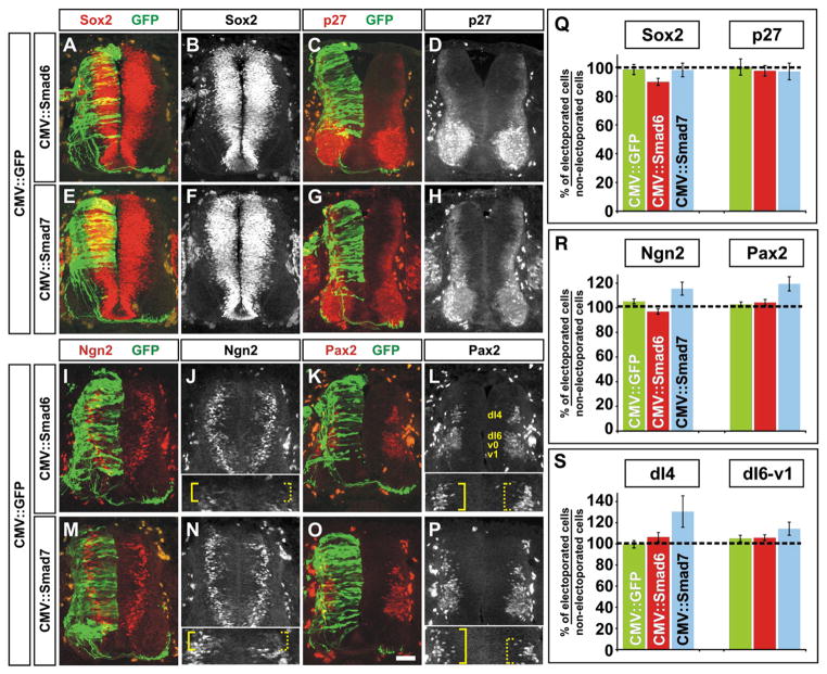
Fig. 4.
Smad7, but not Smad6, mis-expression leads to a compensatory increase in the number of dI4 neurons. (A–P) GFP (green) in combination with either Smad6 (A–D and I–L) or Smad7 (E–H and M–P) were ectopically expressed throughout the spinal cord under the control of the CMV enhancer at HH stages 14–16. Embryos were harvested at HH stages 24/25 and examined for the number of Sox2+ (A, B, E and F; neural progenitors), p27+ (C, D, G and H; post-mitotic neurons), Ngn2+ (I, J, M and N; dP2–5 progenitors) and Pax2+ (K, L, O and P; post-mitotic dI4 and dI6-v1 neurons) cells in the dorsal half of the spinal cord. The inserts in panels J and N show a magnified view of the most dorsal region of the spinal cord. Ngn2+ cells are present more dorsally only on the Smad7 electroporated side of the spinal cord (compare solid brackets, J and N). The inserts in panels L and P show a magnified view of the dI4 population of Pax2+ neurons. Pax2+ cells were present in a more dorsal location only after Smad7 mis-expression (compare solid brackets, L and P). (Q) There was no significant difference in the number of Sox2+ or p27+ cells following electroporation of either CMV::Smad6 (Student’s t-test, Sox2: p>0.18, n=19 sections taken from 5 embryos; p27: p>0.45, n=19 sections taken from 4 embryos) or CMV::Smad7 (Sox2: p>0.59, n=18 sections taken from 4 embryos ; p27: p>0.76, n=13 sections taken from 4 embryos) compared to the CMV::GFP+ control (Sox2: n=20 sections taken from 3 embryos; p27: n=19 sections taken from 3 embryos). (R) In contrast, there was a significant increase in the number of both Ngn2+ and Pax2+ cells following electroporation of CMV::Smad7 (Student’s t-test, Ngn2: p<0.027, n=26 sections taken from 7 embryos; Pax2: p<0.0012, n=33 sections taken from 7 embryos) but not CMV::Smad6 (Ngn2: p>0.13, n=51 sections taken from 11 embryos; Pax2: p>0.61, n=68 sections taken from 11 embryos) compared to the CMV::GFP+ control (Ngn2: n=54 sections taken from 8 embryos; Pax2: n=66 sections taken from 6 embryos). (S) Further examination of the Pax2+ population showed that the mis-expression of Smad7 resulted in significantly more neurons in the more dorsal dI4 Pax2+ cell population compared to the more ventral dI6-v1 Pax2+ population (dI4: p<0.0014 probability of similarity with GFP+ control, n=24 sections from 7 embryos; dI6: p>0.13, n=26 sections from 7 embryos). Scale bar: 25 μm.

