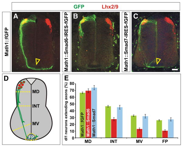
Fig. 5.
Mis-expression of Smad6, but not Smad7, resulted in decreased dI1 commissural axon outgrowth. (A) Lhx2/9+ dI1 commissural neurons (red) electroporated with farnesylated (f) GFP under the control of the Math1 enhancer (Math1::fGFP) at HH stage 15, extend GFP+ axons (green) normally to the floor plate (FP, arrowhead) by HH stage 23. (B) In contrast, Lhx2/9+ dI1 neurons (red) expressing a Smad6-IRES-fGFP cassette (green) have dramatically reduced axon outgrowth by HH stage 23. (C) Lhx2/9+ dI1 axons electroporated with a Smad7-IRES-fGFP cassette (green) extend normally to the FP (arrowhead) by HH stage 23. (D) The extent of the dI1 axon outgrowth was quantified by determining whether dI1 axons had crossed one of four arbitrary lines in the spinal cord: mid-dorsal (MD), intermediate (INT), mid-ventral (MV) or the FP. (E) By HH stage 23, 65–75% of Lhx2/9+ neurons electroporated with GFP, Smad6-IRES-fGFP or Smad7-IRES-fGFP had extended GFP+ axons. Over 35% of these axons had reached the FP in both the GFP (n=465 sections, taken from 10 embryos) or Smad7 (n=116 sections taken from 5 embryos) mis-expression experiments (probability of similarity, p>0.57). In contrast, only 15% of Lhx2/9+ neurons electroporated with Smad6-IRES-fGFP (n=105 sections, taken from 5 embryos) had extended to axons to the FP (probability of similarity to GFP control, p<9.4×10⁻¹¹). Scale bar: 25 μm.

