
Fig. 4. Ad transduces the majority of major prostatic cell types following intraprostate injection.
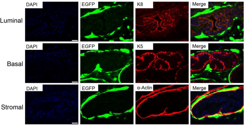
Representative images of immunofluorescence staining of prostate luminal cells (top panel), basal cells (middle panel) and stromal cells (bottom panel), marked by K8, K5 and α-actin staining, respectively.