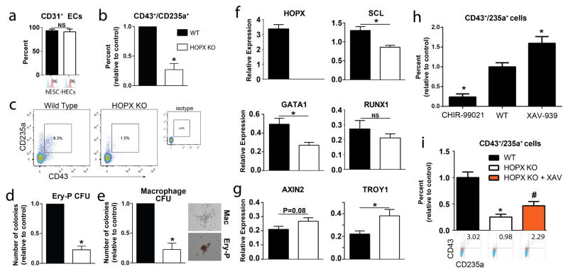
Figure 6. HOPX is functionally required for primitive hematopoiesis.
(a) Fate specification of CD31 endothelial cells is not effected by HOPX KO. (b) HOPX KO does not impact functional endothelial lumen formation assays in collagen. Scale bars = 200 μm. (c–e) Hematopoiesis analysis of CD43+/CD235a+ derivatives in day 5 H-ECs with representative FACS plots (c) and colony forming assays in methylcellulose assaying for Ery-P (d) and macrophage CFUs (e) in WT vs. HOPX KO cells. Representative CFU images are shown to the right for primitive erythroid (Ery-P), and macrophage (Mac) colonies. (f–g) Quantitative RT-PCR analysis of HOPX and genes involved in hematoendothelial cell fate specification (SCL) and function (GATA1 and RUNX1) (f) as well as Wnt/β-catenin target genes AXIN2 and TROY1 in WT vs. HOPX KO cells (g). (h) Wnt-dependent regulation of CD43+/CD235a+ primitive hematopoiesis assessed by treatment of cells with the Wnt agonist CHIR-99021 (1 μM) or the Wnt antagonist XAV-939 (1 μM). (i) Differentiation of HOPX KO and WT cells into H-ECs showing that addition of the Wnt inhibitor XAV-939 (1 μM) partially rescues hematopoietic deficiencies observed in HOPX KO cells. n=4–8 per group. Values are presented as mean ± sem. *P<0.05 vs. WT. # P<0.05 vs. HOPX KO cells.

