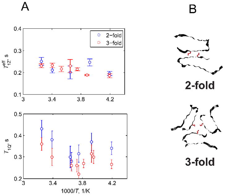
Figure 19.
Plots of and T1Q vs. 1000/T at 17.6 T magnetic field strength for the M35 site in the amyloid fibrils comprised of amyloid-β (1–40) protein in the two-fold and three-fold symmetric polymorphs. The measurements employed the QCPMG signal-enhancement scheme. B) Structural representation of the 2-fold and 3-fold symmetric polymorphs of amyloid-β fibrils, view down the fibril axis.(20, 67) The M35 side-chain pointing into the water-accessible cavity is shown in red. Reprinted with permission from (66), Copyright (2016) Elsevier, License 3995621063676.

