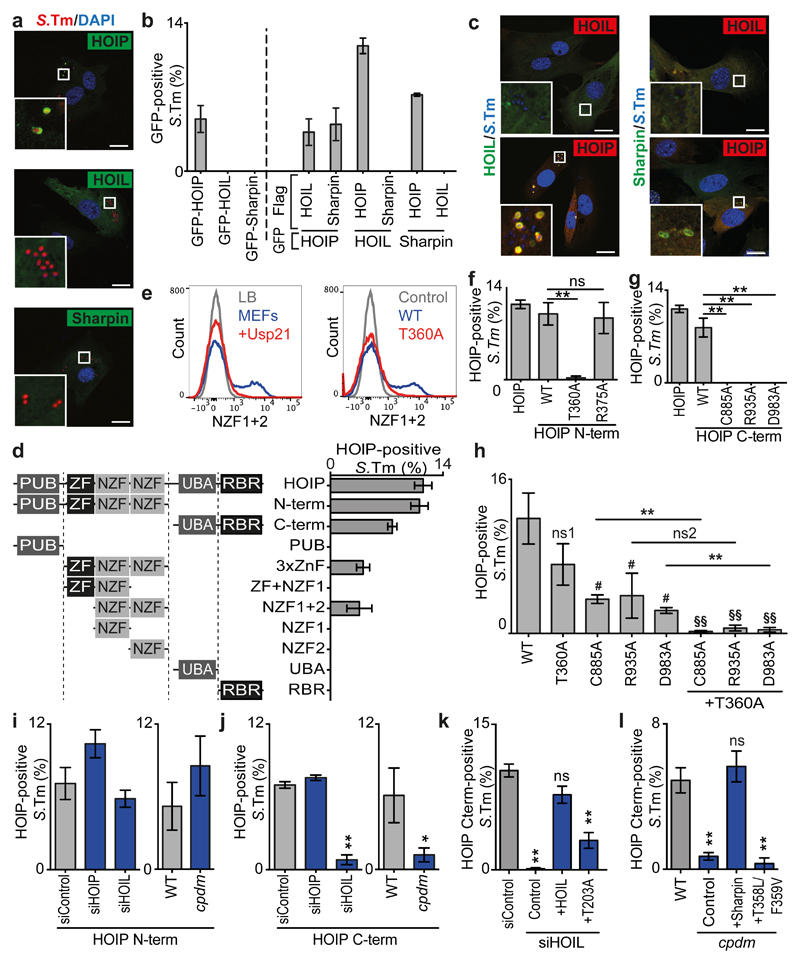
Fig.2. HOIP senses and amplifies the ubiquitin coat of S.Typhimurium.
(a,c) Confocal micrographs of PFA-fixed MEFs infected with (a) mCherry-expressing or (c) DAPI-stained S.Typhimurium (blue) and expressing (a) GFP-tagged LUBAC subunits only or (c) co-expressing Flag-tagged LUBAC subunits (red) at 1h p.i.. Data are representative of three independent experiments. Scale bar 20μM. Split channels displayed in Fig.S2A,C.
(b,d,f-l) Percentage of marker-positive S. Typhimurium as counted by eye using widefield microscopy at 1h p.i. in PFA-fixed MEFs expressing (b) the indicated GFP- and FLAG-tagged LUBAC subunits or (d,f-l) GFP-tagged HOIP alleles. (i-l) MEFs from wild type or Sharpincpdm mice were treated with control siRNA or siRNA against the indicated murine LUBAC components and complemented with Flag-tagged human HOIP, HOIL-1 or Sharpin alleles as indicated. (b) Mean±SD of triplicate coverslips, representing two independent repeats. (d,f-l) Mean±SEM of triplicate coverslips from three independent repeats, n>100 bacteria per coverslip. *p<0.05, **p<0.01, (f,g,h,l) one-way ANOVA with Dunnett’s multiple comparisons test or (h,j) Student’s t-test. (h) # or ns1; compared to WT, §§; compared to T360A.
(e) Flow cytometry of S.Typhimurium grown in LB as indicated or extracted from MEFs (all other samples), treated with Usp21 as indicated, and stained with recombinant HOIP NZF1+2 (WT, blue), mutant allele (T360A, red) or secondary reagent only (Control, grey).

