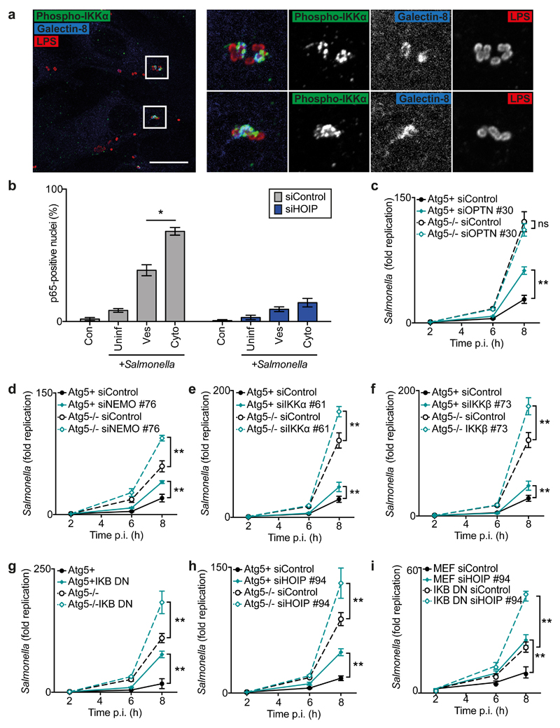
Fig.4. LUBAC activates autophagy and NF-κB, which independently restrict cytosolic S.Typhimurium.
(a) Confocal micrographs of methanol-fixed MEF cells at 1h p.i. with S.Typhimurium, stained with antibodies against LPS (red), phosphorylated IKKα (green) and Galectin-8 (blue). Data are representative of three independent experiments. Scale bar, 20μM.
(b) Percentage of p65-positive nuclei as counted by eye using widefield microscopy at 1h p.i. in siRNA-treated PFA-fixed MEFs infected with GFP-expressing S.Typhimurium and stained for p65 and Galectin-8. Cells in infected sample (+Salmonella) were classified by bacterial status: no bacteria (Uninfected, ‘Uninf’), Galectin-8-ve bacteria only (Vesicular, ‘Ves’) or ≥1 Galectin-8+ve bacteria (Cytosolic, ‘Cyto’). Mean±SEM of triplicate coverslips from three independent repeats. n>100 bacteria counted per coverslip.
(c-i) Fold replication of S.Typhimurium in MEFs treated with indicated siRNAs against (c) Optn, (d) Nemo, (e) IKKα, (f) IKKβ, (h-i) HOIP or (g,i) expressing I-κBαSS32,36AA (I-κBα DN). Bacteria were counted based on their ability to form colonies on agar plates. Mean±S.D. of triplicate MEF cultures and duplicate colony counts, representing two (e) or three (c-d,f-i) independent repeats. **p<0.01, Student’s t-test, (c-i) calculated for 8h timepoint.

