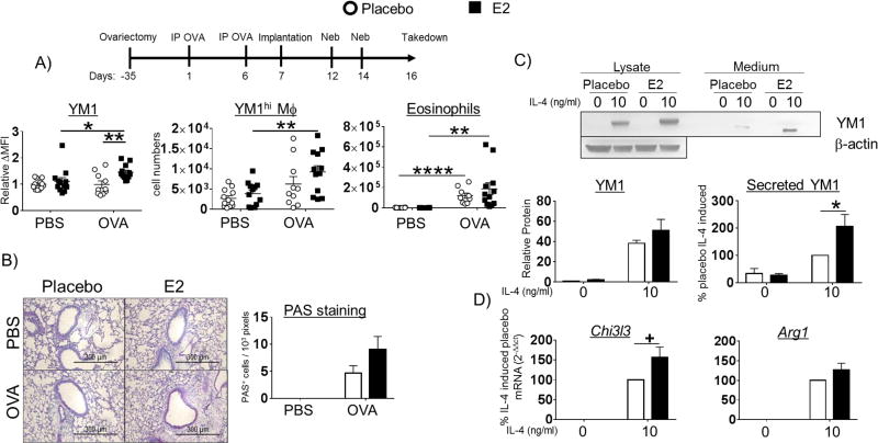
Figure 5. Estrogen enhances M2-polarization in ovariectomized mice.
(A) C57BL/6 mice underwent ovariectomy surgery at 3 weeks of age. The standard OVA protocol was initiated at 8 weeks of age. One day after the second IP delivery of OVA, the mice were implanted with pellets that secrete either placebo or E2 and were then challenged with aerosol OVA. Expression of YM1 ΔMFI (MFI target - MFI isotype) in LIVE/DEAD−CD11c+Siglec F+CD45+ alveolar macrophages normalized to placebo-implanted PBS group and absolute numbers of YM1hi alveolar macrophages and CD11c−Siglec F+CD45+ eosinophils are reported above (upper panels). N = 3 independent experiments with 4 mice / group; *p < 0.05; ** p <0.01 (B) PAS staining was carried out as in Figure 1 A. Representative PAS stained alveoli are shown above (left panel). (C) BMM from pellet E2- and placebo-implanted OVx mice were stimulated with IL-4 for 48h. Protein lysates and culture medium were collected and YM1 expression and secretion was measured by western blot. YM1 in cell lysates was normalized to β-actin, AU = arbitrary unit. (D) Expression of mRNA for Chi3l3, Retnla, and Arg1 in BMMs from placebo- or E2-implanted mice stimulated with IL-4 were measured by qPCR. The data is reported as % placebo IL-4-induced; all samples are normalized to the placebo group stimulated with IL-4 and then multiplied by 100. A representative blot (upper panel) and quantified band intensities are graphed (lower panels). N = 3 independent experiments; *p < 0.05, +p = 0.0705.

