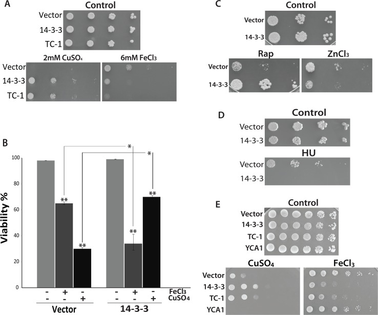
Fig 1. The effects of iron, copper, and HU on cells expressing the anti-apoptotic sequence 14-3-3.
(A) Spot assays were used to assess the ability of wild-type cells expressing 14-3-3, and TC-1 to grow when challenged with 2mM copper (CuSO4) or 6mM iron (FeCl3). (B) The viability of cells containing empty vector and 14-3-3 expressing strains untreated (-) or treated (+) with 1.4mM copper (CuSO4) or 6mM iron (FeCl3) was determined by examining cells stained with trypan blue. Viability is shown as the mean percentage (%) of the cells that survived after treatment in triplicate experiments that were repeated at least 3 times.*; indicates significant differences between control cells (Vector) cells expressing 14-3-3 that were treated with copper or iron (p <0.001). **; indicates significant differences between control cells (not treated) and cells treated with copper or iron (p <0.001). (C) Spot assay was used to examine cell growth ability of the wild-type cells expressing 14-3-3 on nutrient agar containing 200nM rapamycin (Rap) or 20mM zinc chloride (ZnCl2). (D) Spot assay using nutrient agar containing 200mM Hydroxyurea (HU) was used to assess the growth of cells expressing 14-3-3. (E) Yeast cells harboring empty vector, as well as vector expressing 14-3-3, TC-1 and YCA1 were serially diluted and spotted on YNB media with galactose alone (Control) or with 1.8mM copper (CuSO4) or with 5mM iron (FeCl3). The cells were incubated at 30°C to grow for 3 days.

