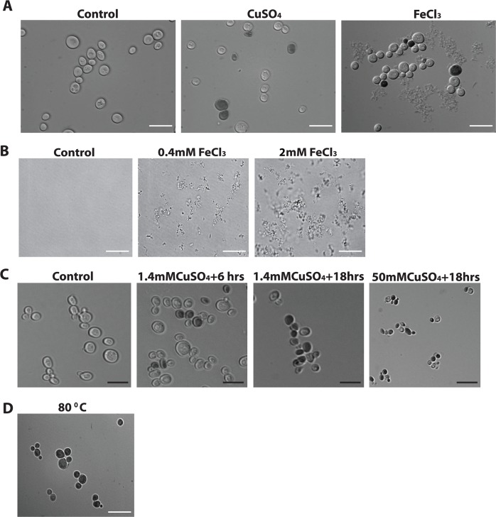
Fig 2. The effect of iron and copper stress on cell morphology.
Yeast cells were grown in glucose for 24 hrs then inoculated in galactose media for 4 hrs followed by the addition of a different stress as explained in A,B and C. (A) Control cells or cells were inoculated with 1.4mM copper (CuSO4) or with 1mM iron (FeCl3) and their morphology were examined following treatment with trypan blue under the microscope. Live cells treated with trypan blue remained clear and appeared blue when dead. (B) Different concentrations of iron (FeCl3) were added to the media in the absence of cells. Media without iron (Control) is clear while with additive iron concentrations, 0.4mM FeCl3 and 2mM FeCl3, precipitation of iron is increased. (C) Yeast cells were non-treated or treated with various intensity of copper concentrations and different exposure time and examined under the microscope after staining with the vital dye trypan blue. Cells were treated with 1.4mM copper (CuSO4) for 6hrs, or for 18 hrs or 50mM copper (CuSO4) for 18hrs. (D) Cells were incubated at 80°C for 20min. and examined after inoculating with trypan blue. White scale bar, 100μm (A, D) and black scale bar, 20μm (B).

