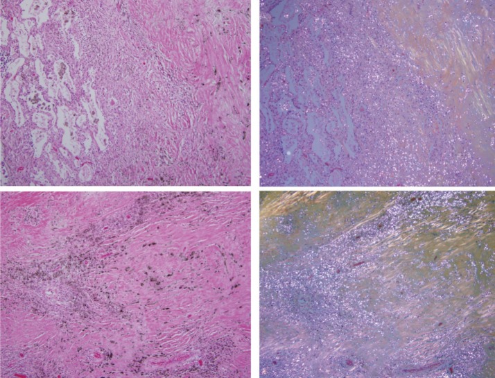
Figure 3.
Coal mine dust desquamative chronic interstitial pneumonia (upper panel, left side of the photomicrograph) in a continuous gradual progression to coal mine dust-related diffuse fibrosis (upper panel, right side of the photomicrograph). Lower panels show coal mine dust-related diffuse fibrosis. Collagen fibers are more or less parallel to each other. On the left side are photomicrographs of H&E-stained tissue (original magnification 100×); on the right side are the same frames under polarized light.

