
Figure 2. A HSD drives exacerbates histopathologic inflammation in colons of Il10−/− mice.
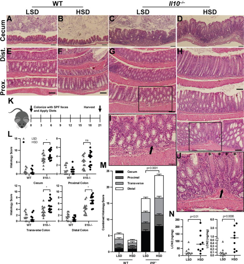
GF mice were colonized with SPF fecal microbiota and harvested 21d later. (A–D) 10× magnification of representative H&E stained sections of cecal tissue from WT (A,B) and Il10−/− (C,D) animals. (E–H) 10× magnification of representative H&E stained sections of colon Swiss rolls, oriented with more proximal tissue on the bottom and distal tissue on the top. Boxes indicate the location of the insert. (I–J) 20× magnifications of boxed inserts in (G,H) respectively. Arrows indicate mononuclear cell infiltration; asterisks indicate crypts where goblet cells have expelled mucus, thus creating an apparent ‘loss’ of goblet cells. Sections shown originate from a representative mouse that had histologic scoring at the average histologic score for the group (in L,M). Data are representative of 3 replicate experiments. (K) GF mice were colonized with SPF feces and fed either a HSD or LSD. Animals were harvested after 3 weeks. (L) Histologic scoring of WT and Il10−/− animals for the cecum, proximal colon, transverse colon, and distal colon. (M) Combined histologic scores, representing the sum of scores from the cecum and colonic regional tissues. (N) LCN2 and MPO ELISA results from feces; results were normalized to protein content as determined by BCA assay. P values determined by Mann Whitney nonparametric tests, or ANOVA for multiple comparisons; * represents p <0.05. Data are combined from 3 (L–M) or 2 (N) replicate experiments.