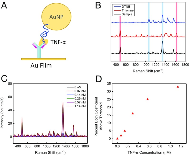
Fig. 5.
Detection of TNF-α using an antibody–aptamer reagent pair. (A) Schematic of the TNF-α detection system, in which labeled antibodies are immobilized on the AuNP film and TNF-α aptamers (blue line) are immobilized on AuNPs. (B) The AuNPs and gold films were respectively labeled with the Raman tags thionine and DTNB. (C) As the concentration of TNF-α protein increases in the solution, an increase in Raman intensity of both labels is observed. (D) Standard TNF-α binding curve in a background of 5 µM albumin.

