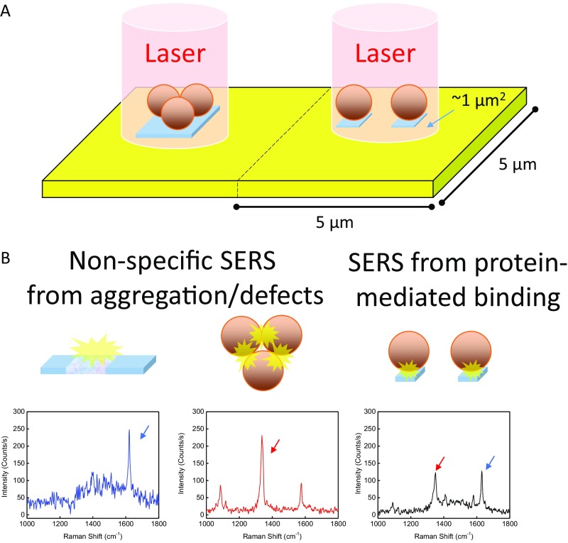
Fig. S1.
Potential sources of SERS hotspots. (A) The center of each 25-μm2 area is interrogated by a 1-μm2 focused laser beam. (B) Three types of hot spots are possible in the assay: (Left) false positives originating from defects in the film, which are dominated by the MB Raman label, with a strong peak at 1,630 cm−1; (Center) false positives from AuNP aggregates, which are dominated by the NBT Raman label with a strong peak at 1,337 cm−1; and (Right) true-positive protein-mediated binding, indicated by near-equal enhancement of MB and NBT peaks.

