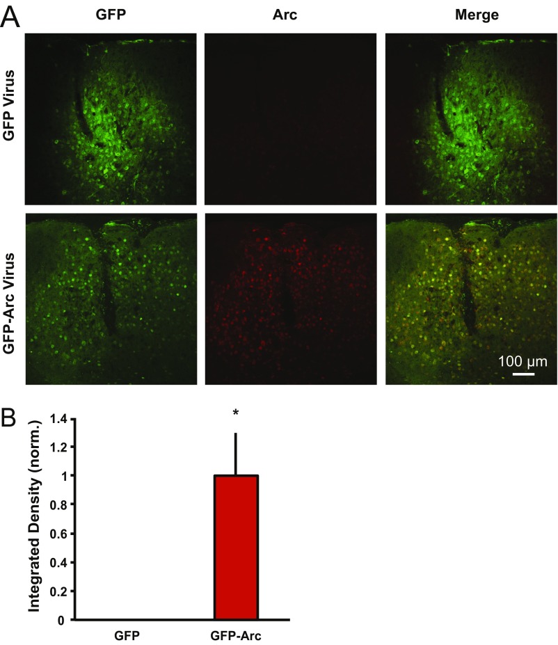
Fig. S3.
GFP-Arc lentivirus increases Arc expression in P180 WT visual cortex. P180 WT mice were injected unilaterally in the visual cortex with lentivirus expressing either GFP alone or GFP-Arc. (A) Representative images from visual cortex of a mouse injected with GFP lentivirus (Upper) and a mouse injected with GFP-Arc lentivirus (Lower). (Left and Middle) GFP expression from GFP and GFP-Arc lentiviruses (no antibody staining) is shown. (Right) IHC for Arc expression is shown. (B) Quantification of Arc expression from GFP- and GFP-Arc–injected mice (four GFP-injected, four GFP-Arc–injected). When images were set to a threshold determined by the maximum Arc expression in GFP-Arc–injected mice, there was no detectable Arc expression in layer IV of GFP-injected mice above background staining, as seen in noninjected P180 WT (Fig. 2). However, there was a significant increase in Arc expression in GFP-Arc–injected mice (P = 0.02, Student t test). Data are normalized (norm.) to GFP and displayed as mean ± SEM.

