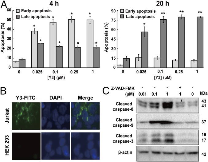
Fig. 2.
Y3 induced the apoptosis of Jurkat cells. (A) Annexin V and 7-AAD staining quantitated cell populations in early and late apoptosis after treatment with serial concentrations of Y3 for 4 and 20 h. Data are shown as the means ± SD (n = 3). Significant differences between control (0 µM) and treatments are shown (*P < 0.05; **P < 0.01). (B) Y3-FITC showed strong binding to the cell surface of Jurkat cells while minimal-to-no binding to HEK 293 in fluorescence microscopy images. (C) Caspases-3, 8, and 9 in Jurkat cells were activated by Y3 as indicated by Western blotting analysis using specific antibodies. Jurkat cells were treated with different concentrations of Y3 and pan-caspase inhibitor Z-VAD-FMK (20 µM) for 20 h.

