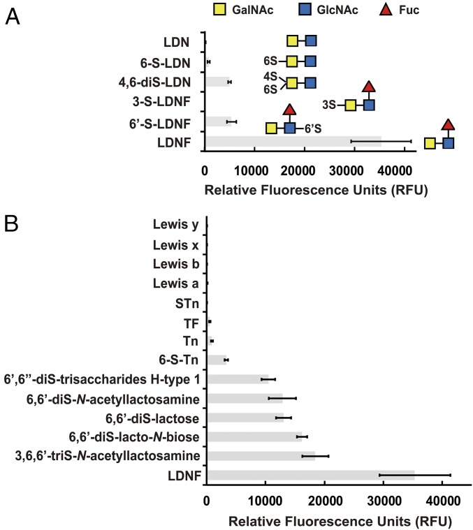
Fig. 3.
Glycan array screening identified LDNF as a specific ligand of Y3. (A) Y3 showed varying binding to glycans of the LDL family with LDNF as the best ligand. Blue square: GlcNAc; yellow square: GalNAc; red triangle: Fuc; S: sulfo. (B) Fluorescence signals of other top glycans and common human antigens in binding to Y3. Figures were generated from data with Y3 at 50 µg/mL in the screening. Data are presented as mean ± SD (n = 6).

