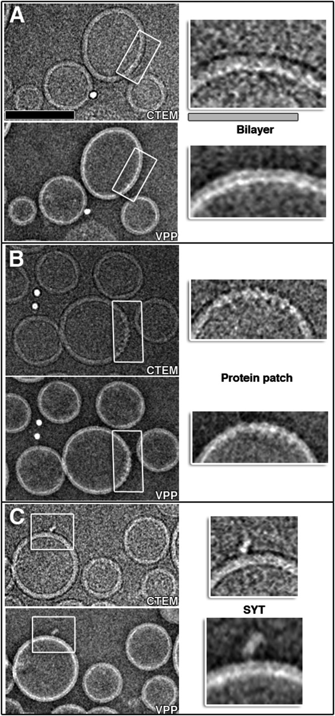
Fig. S2.
VPP cryo-EM. (A–C, Top Left) Conventional cryo-TEM images of a mixture of SV + PM vesicles collected at ∼6.5 μm defocus. (Lower Left) The TEM images of the same mixture of SV + PM vesicles that were collected with VPP at 0 μm defocus. Increased resolvability of the images at focus allowed improved interpretation of the images, as is shown in the close-up views of the areas indicated by gray boxes in the Right Top and Bottom panels showing vesicle bilayers, protein patches, and protruding membrane proteins, such as Syt1. (Scale bars: black, 100 nm; gray, 25 nm.) All other tomograms throughout this work were collected with VPP. Tomogram 2D slice thickness: 1 pixel = 3.42 Å.

