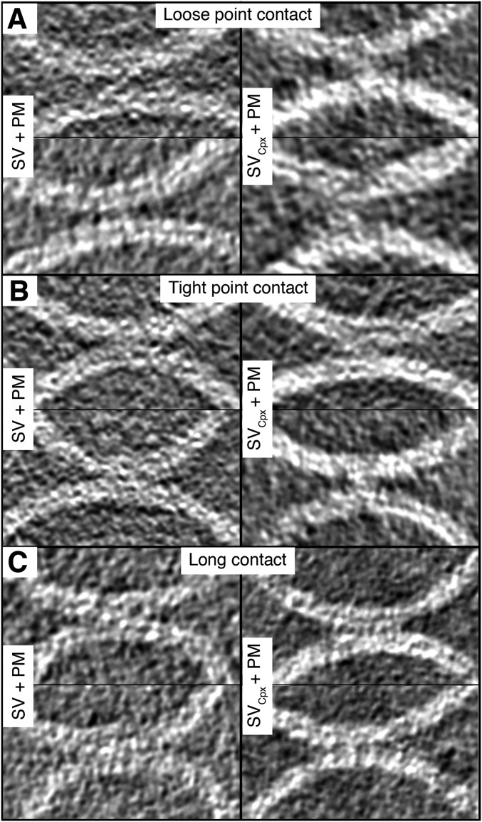
Fig. S4.
Contact sites of SV + PM vesicles with and without Cpx. Representative gray scale tomographic 2D-slice views of contact sites, in addition to those shown in Fig. 2. Contact sites with and without Cpx are shown in the right and left panels, respectively. (A) Loose point contact, (B) tight point contact, and (C) long contact. Tomogram 2D slice thickness: 1 pixel = 3.42 Å.

