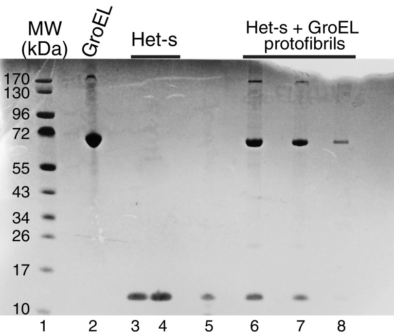
Fig. S2.
Characterization of GroEL, Het-s, and Het-s/GroEL protofibrils by Coomassie-stained SDS/PAGE (4–12% wt/vol). Lane 1, molecular mass standards; lane 2, 10 μM (in subunits) GroEL (subunit theoretical molecular mass = 57.1 kDa); lanes 3 and 4, 10 and 20 μM Het-s(218–289) (theoretical molecular mass = 8.7 kDa), respectively; lane 5, supernatant after spinning down protofibrils obtained 5 min after addition of 100 μM (in subunits) GroEL to 100 μM Het-s; lanes 6–8, pelleted protofibrils (obtained 5 min after addition of 100 μM in subunits GroEL to 100 μM Het-s) dissolved in SDS loading buffer at concentrations of 7, 4, and 2 μM, respectively, in GroEL (subunit concentration) and Het-s in a 1:1 ratio.

