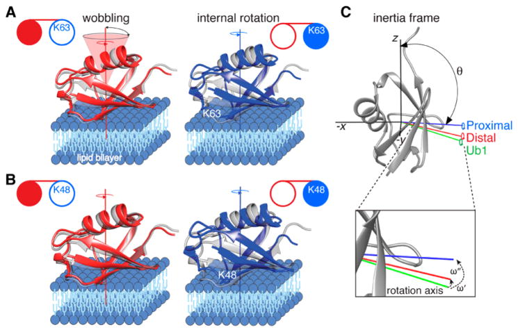
Figure 3.
Internal rotation and wobbling of the proximal and distal domains of Ub2 on the surface of lipid-based nanoparticles. (A) K63- and (B) K48-linked Ub2. The distal and proximal domains of Ub2 are shown as red and blue ribbons, respectively, with Ub1 in gray for comparison. Each domain rotates about an internal axis of rotation, approximately perpendicular to the lipid bilayer, and simultaneously wobbles in a cone centered around this axis. (C) Relationship between the axes (−x, −y, and z) of the inertia tensor (black) in the molecular frame with the internal rotation axes of Ub1 (green) and the distal (red) and proximal (blue) domains of K48-linked Ub2. θ is the angle subtended by each internal rotation axis and the z axis of the intertia tensor; φ (not shown) is the angle subtended by the x axis of the inertia tensor and the projection of the internal rotation axis on the x–y plane. The angle formed between the internal rotation axis of Ub1 (green) and those of the distal (red) and proximal (blue) domains of Ub2 are denoted by ω′ and ω″, respectively. These angles are calculated from the polar angles (θi, φi) defining the position of axis i (i = 1,2) in the inertia frame using the relationship cos(ω) = cos(θ1) cos(θ2) + sin(θ1) sin(θ2)cos(φ1 − φ2). The U-[15N/2H]-labeled domains of Ub2 are shown as filled-in circles in the cartoons in panels A and B.

