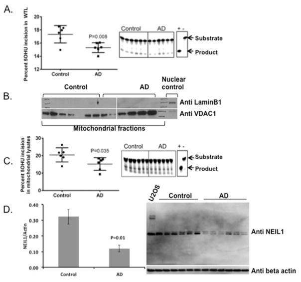
Figure 1.
5OHU incision activity in AD and control extracts. A) Whole tissue lysates from control and AD postmortem brains were assessed for 5OHU incision using 5OHU in a bubble (Table 2). The data are represented by a mean ± standard error, n=6. B) Mitochondrial purity was tested by western analysis of nuclear and mitochondrial markers. C) Mitochondrial lysates from control and AD postmortem brains were assessed for 5OHU incision activity using the same substrate as in panel A containing 5OHU in the bubble. The data are represented by a mean ± standard error, n=6. D) Western analysis of NEIL1 protein in the whole tissue lysates of AD and control samples.

