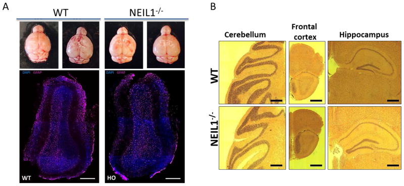
Figure 4.
Histology and immunohistochemistry of the WT and NEIL1−/− mouse brains (A) Top panel represents images of the brains isolated from Neil1+/+ and Neil1−/− 10 months old mice. Bottom panel shows the images (coronal sections) of the adult olfactory bulb stained with DAPI and astrocyte’s marker GFAP (Scale bars, 300 μm). (B) Hematoxylin and eosin staining of various regions from WT and NEIL1−/− mouse brains (Scale bars, 500 μm).

