
Figure 1. Morphological change of SH-SY5Y cells differentiated from mitotic into post-mitotic (neuron-like) cells.
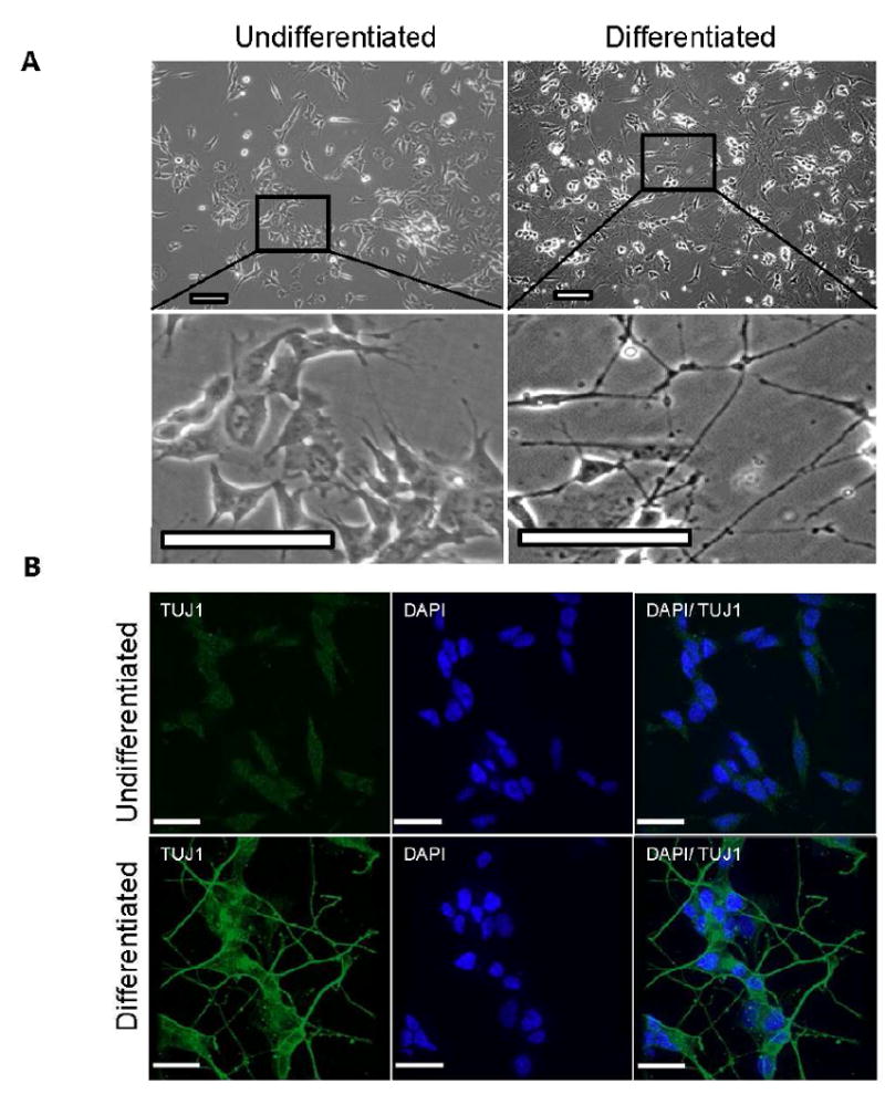
A. (Panels left) Neuroblastoma cells before differentiation. Cells are treated with 10 μM retinoic acid for 5 days; at this time point some cells have extended short neurites but many cells are still dividing. (Panel right) Cells after a further 5 days of culture in serum-free medium containing 10 ng/ml brainderived growth factor (BDNF); at this time point the majority of cells are post-mitotic and exhibit long neurite formation B. The neuron-specific class III beta-tubulin (Tuj1) marker was present in the outgrowth axons of neurons after retinoic acid and BDNF treatment. In comparison untreated SH-SY5Y cells only showed light staining of Tuj1. (Scale bar = 25 μM)