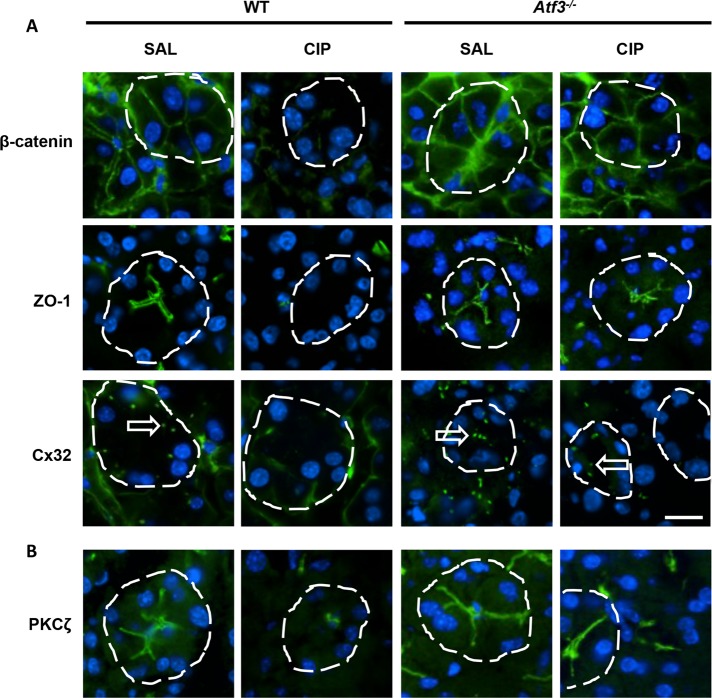
FIGURE 6:
Atf3–/– acinar cells maintain cell junction complexes during CIP. IF for markers of (A) adherens junctions (β-catenin), tight junctions (ZO-1), and gap junctions (Cx32) or (B) cell polarity (PKCζ). Expression and localization is maintained in Atf3–/– acini 4 h into CIP treatment, contrasting the loss of accumulation observed in CIP-treated WT mice. Individual acini are delineated by a dashed line and Cx32 plaques indicated by arrows. Bar, 14 µm.

