FIGURE 2:
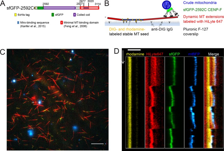
In vitro reconstitution of mitochondria microtubule tracking using CENP-F fragment. (A) Domain organization in recombinant sfGFP-2592C fragment of human CENP-F. Numbers show amino acid position in the full-length human CENP-F. (B) Experimental setup. GMPCPP-stabilized, rhodamine- and DIG-labeled microtubule seeds are immobilized on a coverslip by anti-DIG antibodies. Dynamic microtubules are extended from these seeds by incubation with HiLyte-647–labeled tubulin and GTP. Crude mitochondria expressing a fluorescent marker (mtBFP) are incubated with recombinant sfGFP-2592C and added to the flow chamber. (C) Field of view showing dynamic microtubules (red) growing from coverslip-attached seeds (yellow) and purified BFP-mitochondria (cyan) in the presence of sfGFP-2592C (green). Scale bar, 10 µm. (D) Kymograph showing microtubule seed (yellow), dynamic microtubule extension (red), sfGFP-2592C (green), and a mitochondrial particle (blue). Scale bar, 2 µm (horizontal), 60 s (vertical).

