FIGURE 5:
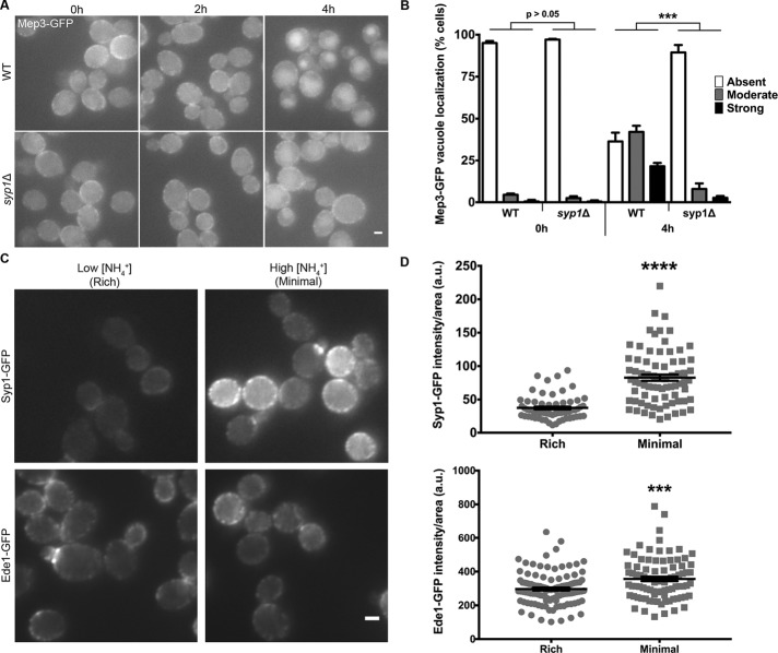
Ammonium-induced trafficking of Mep3 is interrupted in syp1Δ cells. (A) WT and syp1Δ cells were grown to mid-logarithmic phase in rich medium (YPD) and then resuspended in minimal medium (YNB). Cells were imaged every 2 h via live-cell fluorescence microscopy. Scale bar, 2 μm. (B) WT and syp1Δ cells at 0 and 4 h after shift to high ammonium medium were categorized as having strong, moderate, or weak/absent localization of Mep3-GFP to the vacuole (black, gray, and white bars, respectively; ***p < 0.001; each mutant phenotypic class per time point was compared with its respective WT class of the same time point). (C) Cells expressing Syp1-GFP and Ede1-GFP were grown on either rich medium or high ammonium-containing minimal medium and imaged via live-cell fluorescence microscopy. Scale bar, 2 μm. (D) Intensity of Syp1- or Ede1-GFP fluorescence per cell was quantified for each condition; intensity values were corrected for cell size and expressed in arbitrary units (a.u.). Error bars indicate mean ± SEM; ***p < 0.001, ****p < 0.0001 compared with WT).

