Figure 2.
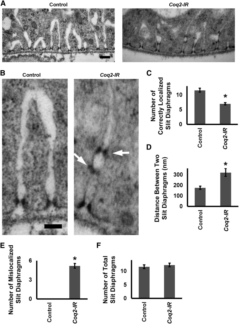
Coq2 gene silencing induced abnormal slit diaphragm localization and collapsed lacunar channels with multiple slit diaphragms (A) TEM showing normal (control) nephrocyte ultrastructure with slit diaphragms and lacunar channels uniformly spaced along the circumference of the cell. Slit diaphragms localized exclusively at the mouth of the channel. In Coq2-IR nephrocytes, channels appeared narrower (collapsed) and interchannel spacing was interrupted and irregular. Slit diaphragms occurred not only at the channel mouth but also ectopically along the interior channel membranes. Scale bar, 200 nm. (B) Higher magnification TEM comparing normal control and Coq2-IR slit diaphragm and lacunar channel ultrastructure. Ectopic slit diaphragms arranged in ladder-like configuration at points of channel narrowing are indicated by arrows. Scale bar, 100 nm. (C) Quantitation of normally localized slit diaphragms in control versus Coq2-IR nephrocytes. Average number of slit diaphragms positioned at mouths of lacunar channels per 2000 nm of cell circumference (*P<0.05). (D) The average distance (in nm) between normally localized slit diaphragms in control versus Coq2-IR nephrocytes (*P<0.05). (E) Quantitation of ectopic slit diaphragms in control versus Coq2-IR nephrocytes. Average number of slit diaphragms positioned along interior channel membranes per 2000 nm of cell circumference (*P<0.05). (F) Quantitation of slit diaphragms (normally localized plus ectopic) in control versus Coq2-IR nephrocytes. Total number of slit diaphragms per 2000 nm of cell circumference (*P<0.05).

