Figure 11.
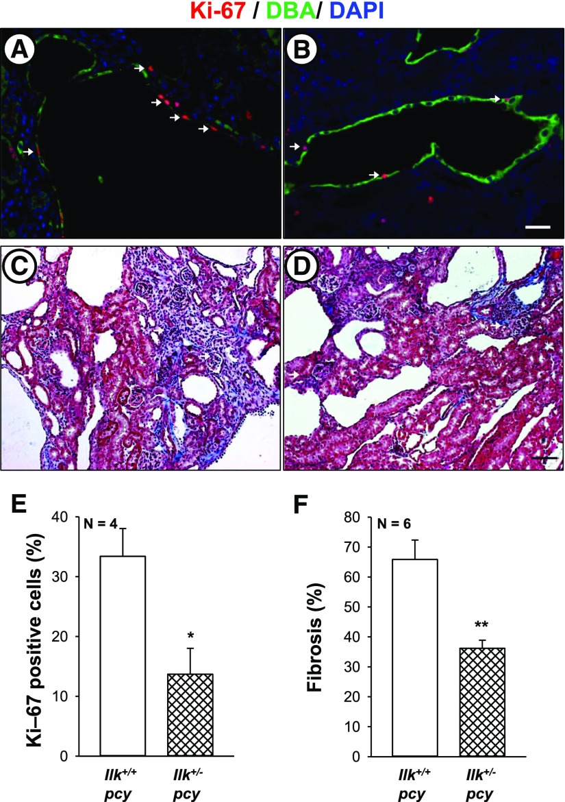
ILK knockdown reduces renal cell proliferation and fibrosis in pcy mice. Proliferating cells were determined in kidney sections from (A) Ilk+/+;pcy/pcy (Ilk+/+ pcy) and (B) Ilkfl/+;Pkhd1-Cre;pcy/pcy (Ilk+/− pcy) mice that were stained for Ki-67 (red), DBA (green), and DAPI (blue). Arrows indicate Ki-67–positive cells. Images (A and B) were taken at the same magnification. Scale bar=5 µm. Representative kidney sections from (C) Ilk+/+ pcy and (D) Ilk+/− pcy mice were stained with Masson trichrome to visualize interstitial fibrosis. Scale bar=50 µm. (E) Comparison of nuclear Ki-67 staining in Ilk+/+ pcy and Ilk+/− pcy sections, normalized to total nuclei. (F) Tissue sections stained with Masson trichrome were coded to conceal the group assignment and visually scored for percentage of interstitial edema and fibrosis. *P<0.05 and **P<0.01, compared with Ilk+/+ pcy.

