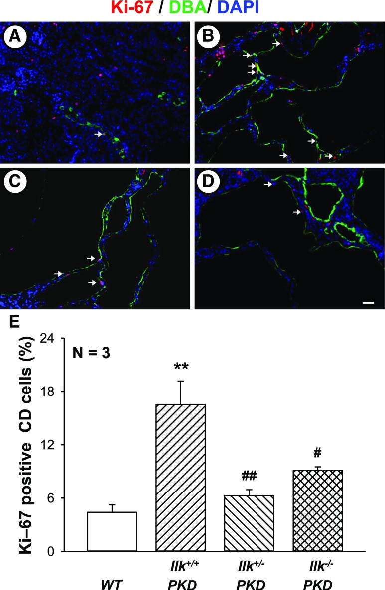
Figure 5.
ILK knockdown reduces renal cell proliferation in PKD mice. Representative kidney sections from (A) WT, (B) Ilk+/+;Pkd1fl/fl;Pkhd1-Cre (Ilk+/+ PKD), (C) Ilkfl/+;Pkd1fl/fl;Pkhd1-Cre (Ilk+/−PKD), and (D) Ilkfl/fl;Pkd1fl/fl;Pkhd1-Cre (Ilk−/− PKD) mice were stained with an antibody to Ki-67, a marker for cell proliferation (red). Tissues were also stained for DBA (green) to detect CDs and the DAPI nuclear stain (blue). Arrows indicate Ki-67–positive CD cells. All images were taken at the same magnification. Scale bar=5 µm. (E) Summary data for the number of cells positive for nuclear Ki-67, normalized to the total nuclei in DBA-positive CDs. Data are mean±SEM for kidneys from three mice per group. **P<0.01 compared with WT, #P<0.05 and ##P<0.01 compared with Ilk+/+ PKD.

