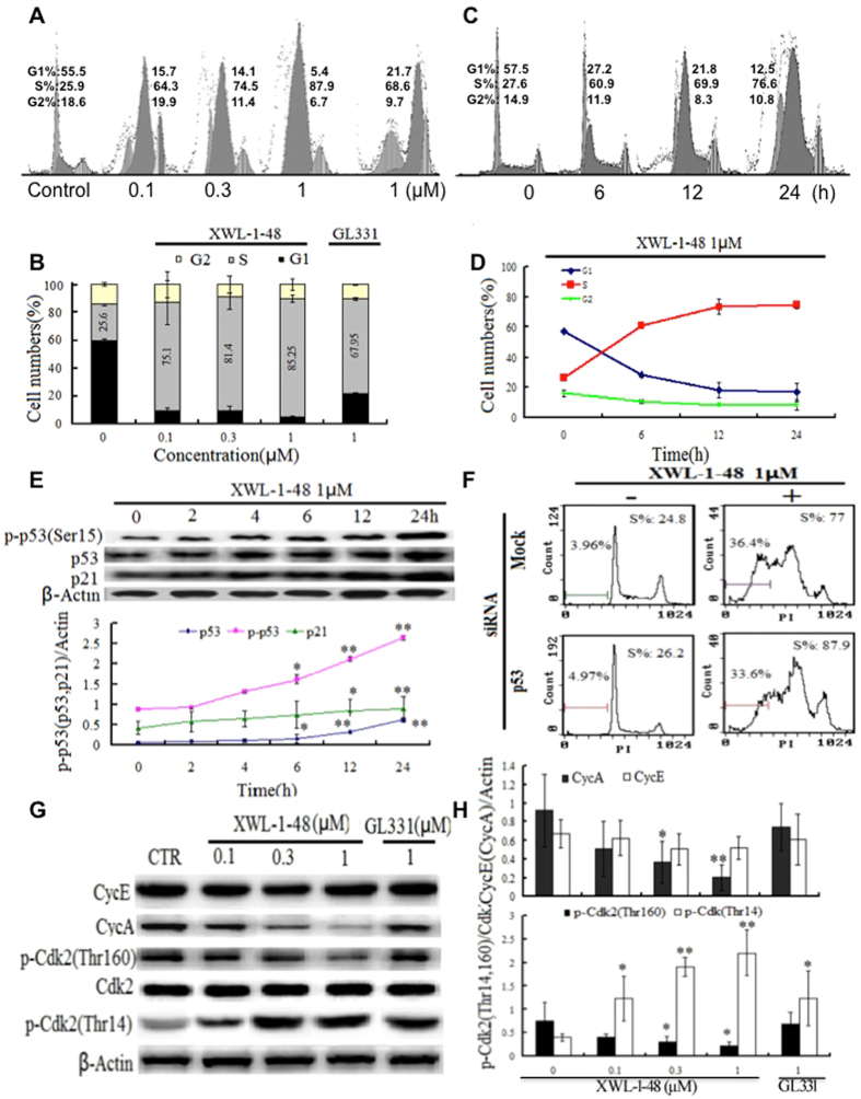
Figure 3.
Effect of XWL-1-48 on cell cycle in HCC. (A) HepG2 cells were treated with XWL-1-48 (0.1, 0.3, 1 µM) and GL331 (1 µM) for 24 h, then cells were harvested, fixed and stained with PI for flow cytometry. (B) Data were shown as mean ± SD of three independent experiments. (C,D) HepG2 cells were incubated with XWL-1-48 (1 µM) for indicated time point, and cell cycle distribution was measured by flow cytometry. (E) HepG2 cells were treated by XWL-1-48 for 0, 2, 4, 6, 12, 24 h. The expression of p-p53 (ser15), p53 and p21 was determined by western blot analysis. A representative of image and curve from three independent experiments is shown. (F) Targeted inhibition of p53 using siRNA for 24 h, further treated by XWL-1-48 for an additional 24 h, then cell cycle were detected by flow cytometry. (G,H) after exposure to XWL-1-48 (0.1, 0.3, 1 µM) and GL331 (1 µM) for 24 h, CycE, CycA, p-CDK2 (thr12, thr160) and CDK were determined by western blot analysis. A representative result of three experiments is shown. *p < 0.05, **p < 0.01 vs. control.

