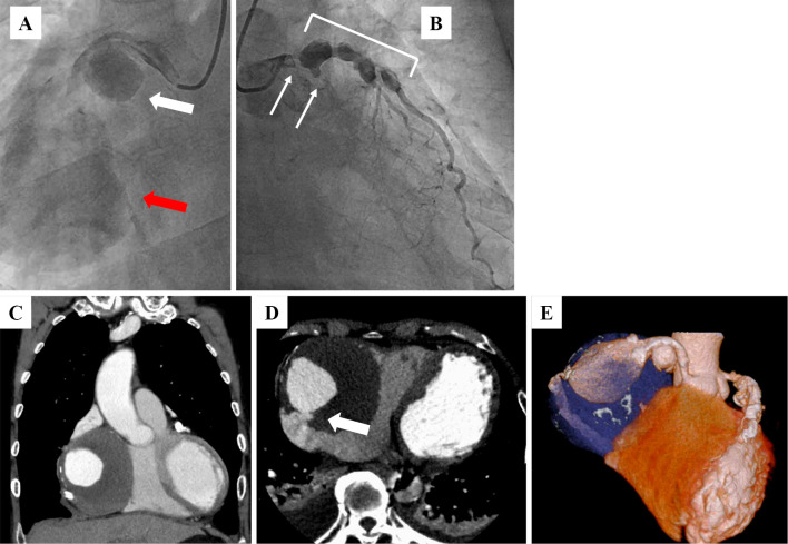
Figure 2.
Emergent coronary angiography showed two giant coronary artery aneurysms (CAAs; A: white and red arrows) in the RCA, severe coronary stenosis (>90%) in the left main trunk (LMT), an occluded left circumflex artery (B: white arrows) and multiple CAAs in the LMT and the proximal portion of the left anterior descending artery (B: white rectangle). Emergent computed tomography showed an 85-mm round mass in the RCA (C-E), which had ruptured into the right atrium (D: white arrow).

