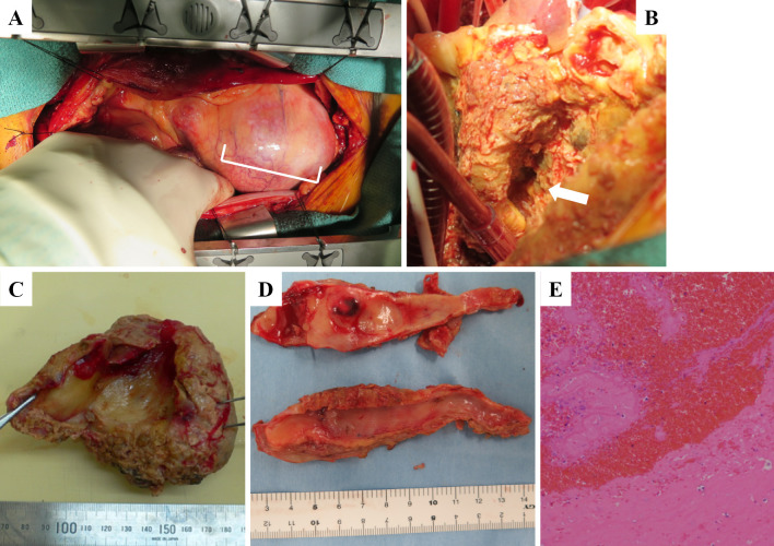
Figure 3.
Intraoperative view of giant coronary artery aneurysm (A: white rectangle). After the giant CAA was removed. we could see the fistula from the coronary artery to the right atrium (B: white arrow). The resected aneurysm was filled with giant organized thrombi and fresh red thrombi (C). We observed severe atherosclerosis in the efferent vessels of the giant CAA (D). A pathological examination revealed organized thrombi and blood cells with no blood vessel components (E).

