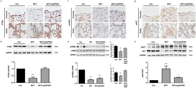
Figure 8.
Effects of PGE1 on PTEN expression in the proximal and distal pulmonary arteries of rat lungs. Immunohistochemistry shows (a) PTEN, (c) pCREB and (g) pAKT expression in the proximal (scale bar: 50 μm) and distal (scale bar: 25 μm) pulmonary arteries. PTEN, pCREB and pAKT levels in the medial layer of the pulmonary arteries of the control lungs were identified by brown-colored staining. Representative immunoblotting and densitometric quantification of protein expression in the indicated three groups. (b) PTEN was detected as a 54-kDa band; (d) pCREB was detected as a 43-kDa band and (h) pAKT was detected as a 60-kDa band. (e) The nuclear pCREB and (f) the intensity of pCREB was detected as a brown staining. The PTEN and pCREB expression levels were decreased but pAKT expression was increased in the pulmonary arteries of the MCT rats. PTEN was elicited and pAKT was diminished in the pulmonary arteries of the MCT rats administered lipid/PGE1. The bars represent the mean ± SEM for n = 3 samples. **P < 0.01 compared with the control (Con) group.

