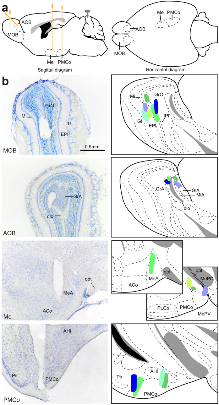
Figure 1.
(a) Schematic diagram of the recording sites, sagittal diagram (left) and horizontal diagram (right). (b) Verification of the electrodes position. The actual recording site (electrode tip) should be along the trace of the electrode. Right, histological verification of the four electrodes placed on the same animal. Left, schematic drawings of the recording regions on all the mice; each colour indicates a different animal. Abbreviations: ACo, anterior cortical amygdaloid nucleus; AHi, amygdalo-hippocampal transition zone; AOB, accessory olfactory nucleus; dlo, olfactory tract, dorsal part; EPl, external plexiform layer of the MOB; Gl, glomerular layer of the MOB; GlA, glomerular layer of the AOB; GrA, granular layer of the AOB; GrO, granular layer of the MOB; IPl, internal plexiform layer of the MOB; Me, medial amygdaloid nucleus; MeA, medial amygdaloid nucleus, anterior subdivision; MePD, medial amygdaloid nucleus, posterodorsal subdivision; MePV, medial amygdaloid nucleus, posteroventral subdivision; Mi, mitral layer of the MOB; MiA, mitral layer of the AOB; MOB, main olfactory bulb; opt, optic tract; PLCo, posterolateral cortical amygdaloid nucleus; PMCo, posterimedial cortical amygdaloid nucleus.

