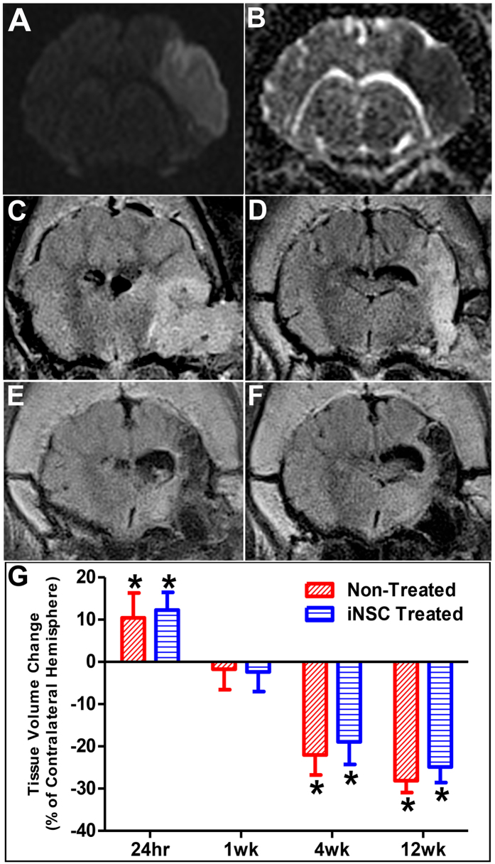
Figure 2.
Ischemic Stroke Results in Significant Brain Swelling and Atrophy. MRI was performed 24 hours post-stroke and 1, 4, and 12 weeks post-transplant. Diffusion weighted imaging (DWI) demonstrated abnormal territorial hyperintensity in the right cerebral hemisphere 24 hours post-stroke (A). Corresponding ADC maps showed hypointense signal at the lesion site, validating the presence of ischemic stroke (B). T2-weighed FLAIR images displayed initial brain swelling in the ipsilateral hemisphere at 24 hours post-stroke (C) that subsides by 1 week post-transplant (D). T2-weighted FLAIR images taken at 4 weeks (E) and 12 weeks (F) post-transplant showed significant brain atrophy at the area of the infarct. Volumetric measurements on T1-weighted images supported these changes (G). Data are expressed as mean ± s.d. Data is generated from scans of 4 animals per treatment group. *Indicates significance from the volume of the contralateral hemisphere (p < 0.005).

