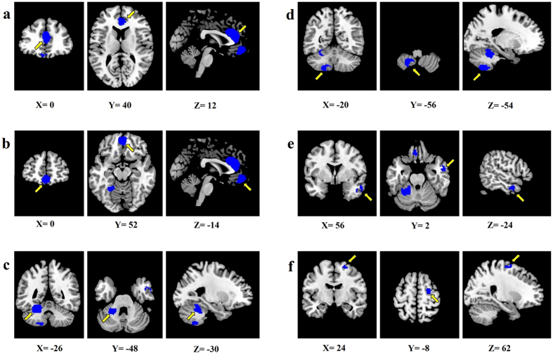
Figure 2.
The meta-analysis revealed six brain regions showing reduced gray matter in patients with OSA compared with that in healthy controls. (a) Bilateral ACG/ApCG; (b) Bilateral SFG (medial rostral part); (c) Left cerebellum, hemispheric lobules IV/V; (d) Left cerebellum, hemispheric lobule VIII; (e) Right MTG; (f) right premotor cortex. Abbreviations: ACG/ApCG, anterior cingulate/paracingulate gyri; MTG, middle temporal gyrus; SFG, superior frontal gyrus.

