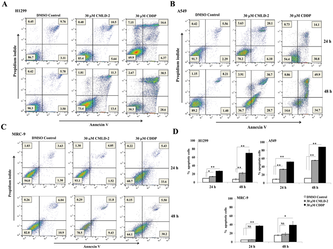
Figure 7.

Annexin V staining demonstrate CMLD-2 treatment induces apoptosis. Flow cytometric analysis shows the percentage of apoptotic and necrotic cells (Q1: necrotic; Q2: late apoptotic; Q3: early apoptotic; Q4: Live cells) at 24 h and 48 h in CMLD-2 -treated cells. (A) H1299; (B) A549; (C) MRC-9. Induction of apoptosis was greater in CMLD-2 -treated H1299 and A549 cells than in CMLD-2 -treated MRC-9 cells. (D) Bar graphs represent the percentage of apoptotic cells at 24 h and 48 h after CMLD-2 treatment. Cells treated with cisplatin (CDDP) served as positive control for each cell line. Error bar denotes SD; NS not significant; *p < 0.05; **p < 0.001.
