Figure 5.
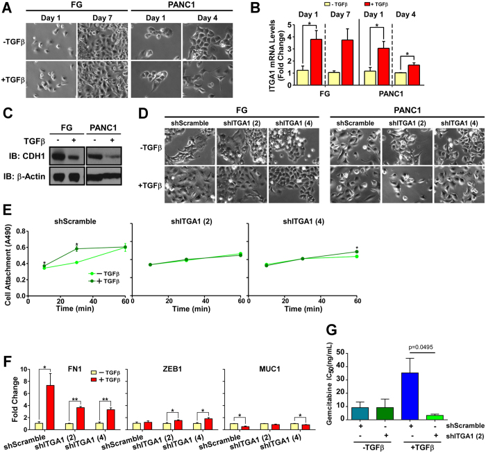
ITGA1 mediates TGFβ/collagen-induced EMT and gemcitabine resistance in PDAC cells. (A) Phase-contrast microscopy images of FG and PANC1 cells plated on collagen (5 μg/mL) and treated with TGFβ. (B) qPCR for ITGA1 in FG cells – 1 and 7 days post-TGFβ treatment, and in PANC1 – 1 and 4 days post-TGFβ treatment. POLR2A was used as the house-keeping gene. (C) Western Blot for CDH1 levels following 7 (FG) or 4 (PANC1) day treatment with TGFβ. Original blot images are cropped to show indicated bands. (D) Phase-contrast microscopy of transduced FG and PANC1 lines with or without TGFβ on day 7 and day 4, respectively. (E) A modified cell viability assay was used to detect attachment of PANC1 transduced lines with or without TGFβ treatment at indicated time points after plating onto collagen (5 μg/mL). (F) qPCR for FN1, MUC1 and ZEB1 expression in transduced PANC1 cells following 4 days of control or TGFβ treatment. (G) IC50 values from gemcitabine dose-response curves of FG shRNA cells on collagen (5 μg/mL) and TGFβ treated or untreated. *Indicates t-test derived p-value less than 0.05.

