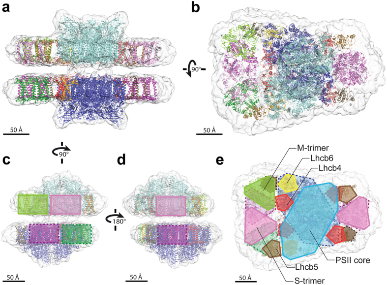
Figure 2.
Fitting the cryo-EM density map of paired C2S2M supercomplexes with high-resolution structures. Side view within the membrane plane (a) and top view towards the luminal surface (b) of the paired C2S2M supercomplexes. The following structures were placed into the cryo-EM map by rigid-body fitting: the PSII dimeric core and monomeric Lhcb4 and Lhcb5 from spinach26 (PDB: 3JCU depleted of subunits PsbP, PsbQ, PsbTn; upper PSII dimer in cyan, lower PSII dimer in blue, upper Lhcb4 in pale red, lower Lhcb4 in dark red, upper Lhcb5 in pale brown, lower Lhcb5 in dark brown), the LHCII trimer from pea36 (PDB: 2BHW; upper S-trimers in pink, lower S-trimers in violet, upper M-trimer in pale green, lower M-trimer in dark green), the predicted structure of monomeric Lhcb6 from pea generated by the PHYRE2 algorithm39 (upper Lhcb6 in yellow, lower Lhcb6 in orange). (c–e) Schematic representations showing the positions of the LHCII trimers (c,d) and of all fitted supercomplex components (e), superimposed on the cryo-EM density map, showing end views within the membrane plane (c,d) and a top view towards the luminal surface (e). Colors match the structures in panels (a,b); solid lines for upper supercomplex components, dashed lines for lower supercomplex components.

